colorectal cancer is among the most common cancers worldwide and a major cause of cancer death.
FOBT, FIT, colonoscopy and pathology
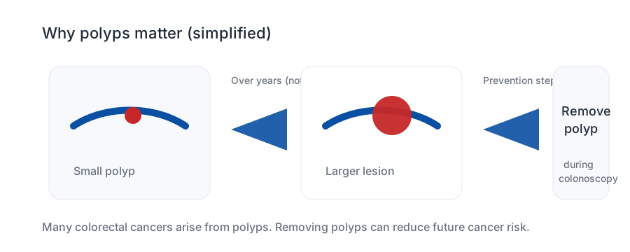
Colon cancer detection works because many cancers start as removable polyps.
A practical guide to warning symptoms, demographics and risks, FOBT/FIT stool testing, stool DNA, colonoscopy, polyp removal, biopsy, histology, genetics, staging scans and the false-positive tradeoffs that come with screening.
is the current US starting age for average-risk screening in many recommendations.
stool blood testing can find hidden bleeding, but a positive test needs colonoscopy.
adenomas and serrated lesions can often be removed before they become cancer.
Brief outline
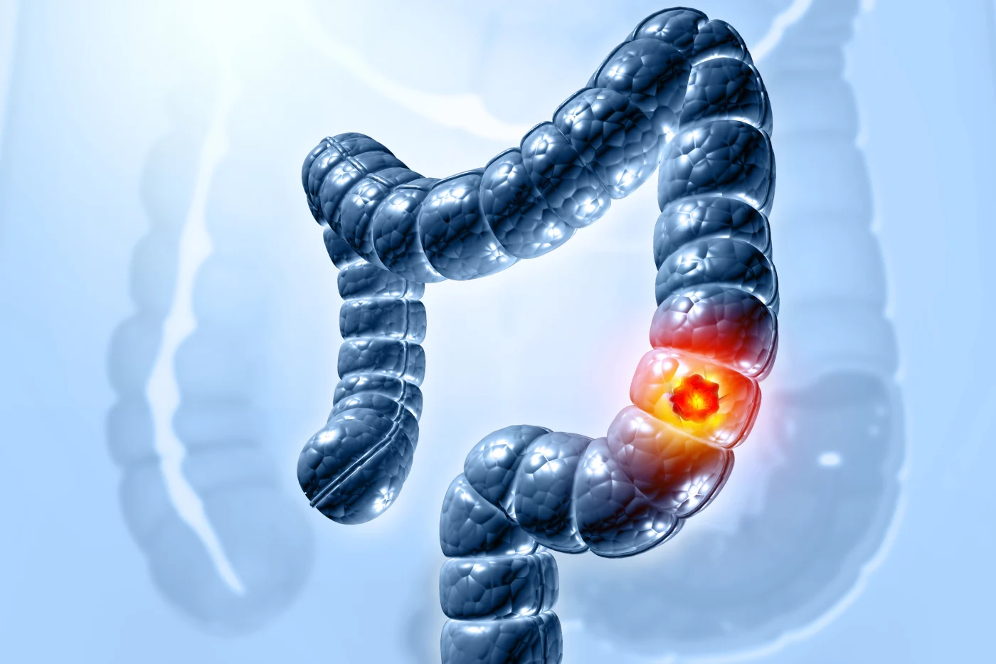
Most colon cancers are adenocarcinomas that arise through a polyp-to-cancer pathway.
Screening is unusually powerful here because it can do two jobs: find early cancer and prevent future cancer by removing precancerous polyps. Rectal cancer overlaps with colon cancer screening, but rectal cancer staging and treatment often add pelvic MRI and neoadjuvant therapy decisions.
Demographics, risks and epidemiology
- Risk rises with age, but early-onset colorectal cancer has increased enough that symptoms in younger adults should not be dismissed.
- Family history, prior adenomas, Lynch syndrome, FAP/MUTYH polyposis, inflammatory bowel disease, smoking, alcohol, obesity, diabetes and processed meat exposure can increase risk.
- Protective patterns include physical activity, healthier weight, not smoking, moderating alcohol and participating in screening.
- Some countries use organized FIT programs; others rely more on opportunistic colonoscopy. The best test is the one that is completed and followed up.
When to start
For average-risk adults, 45 has become the practical US starting age; symptoms at 35 still count.
Age thresholds are for screening people who feel well. They are not rules for ignoring rectal bleeding, iron-deficiency anemia, persistent bowel change, weight loss or unexplained abdominal symptoms in younger adults.
Do not ignore
Symptoms can be vague, so patterns matter.
Not just cancer screening
Bowel symptoms may be IBS, IBD or something else entirely.
A colon cancer detection page should not imply every bowel symptom is cancer. The safer message is that persistent or changing symptoms deserve a diagnosis, not just reassurance or a screening test.
Screening tests
FOBT and FIT are stool blood tests. A positive result is not the endpoint.
FOBT usually means fecal occult blood testing. FIT is fecal immunochemical testing, a more specific stool test for human hemoglobin. Both are screening or triage tools; neither removes polyps or proves where bleeding came from.
gFOBT
Older guaiac fecal occult blood testing can reduce colorectal cancer mortality in organized programs, but diet/drug restrictions and lower specificity are practical limitations.
FIT
FIT is widely used because it is simple, quantitative in many programs and better targeted to lower-GI bleeding. It is usually repeated at set intervals if negative.
Stool DNA-FIT
Multitarget stool DNA plus FIT can detect more cancers and advanced precancerous lesions than FIT alone, but usually with more false positives and colonoscopies.
Colonoscopy
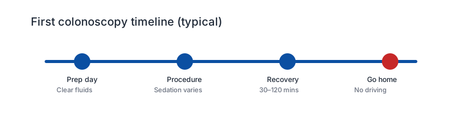
Colonoscopy visualizes the colon, removes many polyps and biopsies suspicious lesions. It is screening, diagnosis and prevention in one procedure.
Positive stool test: do not just repeat it
A positive FOBT/FIT or stool DNA-FIT generally needs diagnostic colonoscopy. Repeating the stool test until it becomes negative can miss intermittent bleeding and delays the examination that can biopsy cancer or remove polyps.
Which stool blood test is better?
For modern population screening, FIT is usually preferred over older guaiac FOBT. FIT is specific for human globin, needs fewer samples, does not usually require diet restrictions, can be quantitative, and lets programs adjust the positivity threshold to match colonoscopy capacity. Guaiac FOBT has the strongest older randomized-trial mortality evidence, but many countries replaced it with FIT because FIT is easier to use and performs better in practice.
| Test type | Specimen | Typical role | Sensitivity / tradeoff |
|---|---|---|---|
| High-sensitivity gFOBT | Stool from 3 bowel movements | Older organized screening approach | CRC sensitivity roughly 62-79% in USPSTF summaries; diet/drug interference and lower convenience than FIT. |
| FIT | Usually one stool sample | Common public-program test | At a 20 microgram Hb/g feces cutoff, pooled CRC sensitivity about 74% and specificity about 94%; advanced adenoma sensitivity is much lower, about 23%. |
| Multitarget stool DNA-FIT | Whole stool sample plus FIT | Private/insurance screening option in some countries | Higher CRC sensitivity, about 93%, and advanced adenoma sensitivity about 43%, but lower specificity, about 85%, meaning more colonoscopies. |
| Blood cfDNA colorectal tests | Blood | Option when stool/endoscopy screening is refused or unavailable | Shield showed about 83% CRC sensitivity and 90% specificity, but only about 13% sensitivity for advanced precancerous lesions, so it is weaker for prevention. |
| Colonoscopy | Direct visualization | Screening, diagnosis, biopsy and prevention | Most complete single test when high quality, but needs bowel prep, sedation planning, capacity and carries small bleeding/perforation risks. |
Free / organized programs
Most public systems choose the cheaper home FIT pathway, then colonoscopy only if positive.
Program rules change and differ by state/province. Symptoms, strong family history, inflammatory bowel disease or known genetic risk should bypass routine screening ages and be assessed clinically.
| Country / region | Current broad public-program pattern | Notes |
|---|---|---|
| United States | USPSTF recommends screening ages 45-75; options include annual FIT, annual high-sensitivity gFOBT, stool DNA-FIT every 1-3 years, CT colonography, sigmoidoscopy or colonoscopy. | Not one national mailed program, but ACA/Medicare coverage usually follows recommended screening. Follow-up colonoscopy after abnormal noninvasive tests is essential. |
| England / NHS | Free FIT kit every 2 years, now being sent to people aged 50-74. | England introduced FIT in 2019 and completed expansion down into the 50s. People over 74 can request kits. |
| Australia | National Bowel Cancer Screening Program: free home test every 2 years for ages 45-74. | From July 2024, ages 45-49 can opt in/order a first kit; ages 50-74 are mailed kits. |
| Canada | Most provinces/territories use FIT or FOBT around ages 50-74 every 1-2 years. | Some jurisdictions are considering or beginning moves toward age 45 because of early-onset colorectal cancer. |
| European Union guidance | Organized population screening is recommended, with FIT generally preferred; many programs target 50-69 or 50-74. | Exact ages and tests vary by country, but biennial quantitative FIT is the main modern direction. |
| New Zealand | Free national bowel screening is being lowered; current public guidance is moving from older 60/58 thresholds toward 56-74 from late September 2026. | Eligibility is publicly funded and staged; symptoms at any age should not wait for a program invitation. |
| Ireland | BowelScreen is a free home FIT program with staged age expansion. | Recent public information describes invitations around ages 57-71, with approved expansion toward younger cohorts. |
Colonoscopy and biopsy
Colonoscopy quality changes what the test means.
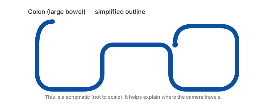
A useful colonoscopy depends on bowel preparation, reaching the cecum, withdrawal time, adenoma detection, complete polyp removal, retrieval for pathology and a clear follow-up interval.
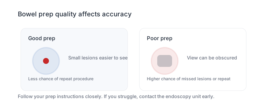
Bowel prep is not a side issue; it is part of the test. In real endoscopy lists, bad prep is common enough to matter. Follow the written instructions exactly, including low-residue diet timing, stopping seeds/iron/fiber if instructed, split-dose laxative timing, clear fluids, and medication advice for diabetes, blood thinners, kidney disease or heart failure. Poor prep can hide flat polyps, small cancers and the cecum itself, and may mean repeating the whole procedure soon rather than waiting years.
The cecum and right colon are a common place for false reassurance to begin. Right-sided cancers may present only as iron-deficiency anemia, and flat sessile serrated lesions can be harder to see than a stalked left-sided polyp. A report should say whether the cecum was reached, whether the appendiceal orifice/ileocecal valve were seen, how good the bowel prep was, what was removed, whether tissue was retrieved and when the next test is due.
You should receive a written colonoscopy report
After the procedure, ask for a copy of the endoscopy report and, if anything was removed or biopsied, the later histology report. The report should make the quality of the examination visible, not just say "normal" or "polyps removed."
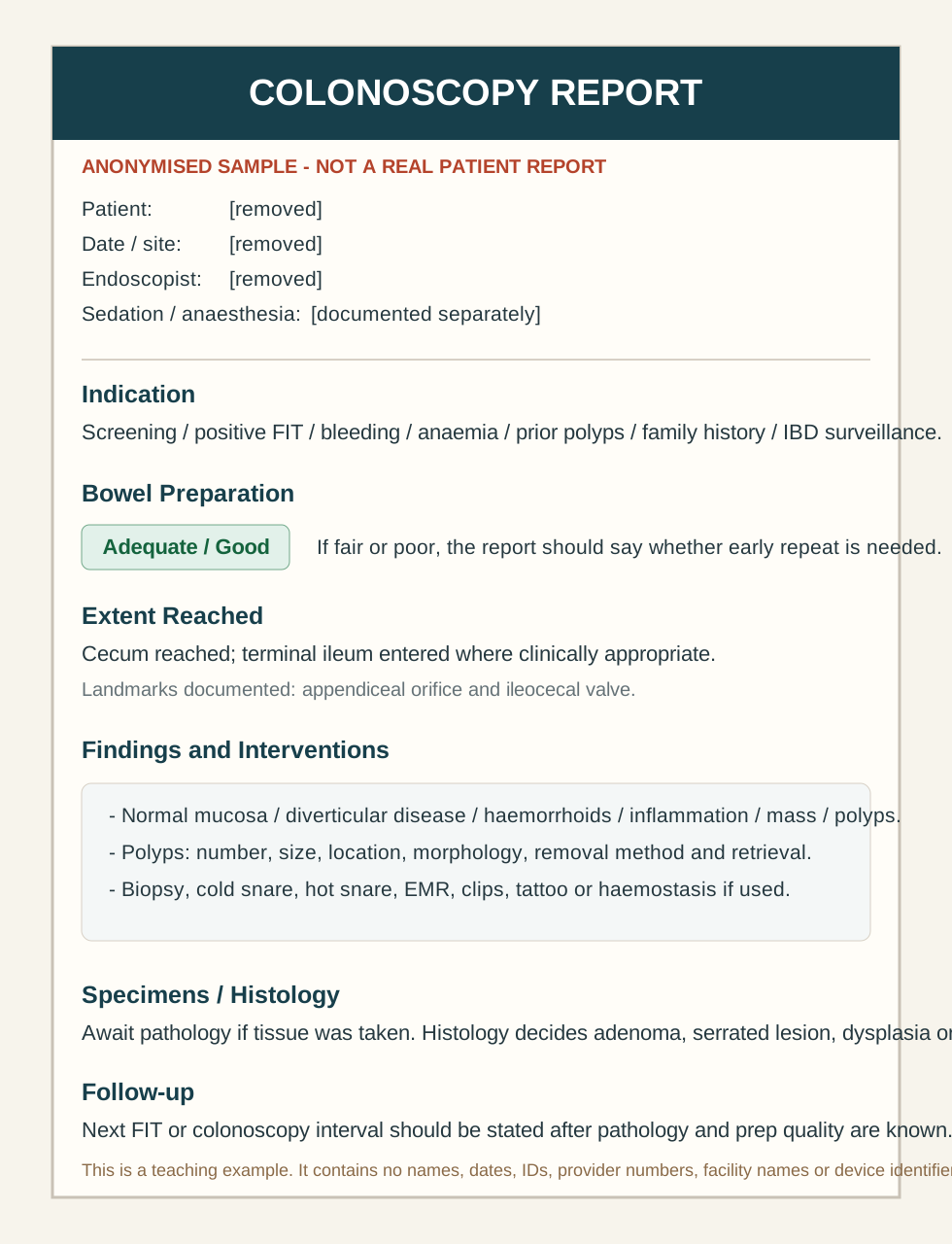
| Example report field | What it should tell you |
|---|---|
| Indication | Why the test was done: screening, positive FIT/FOBT, anemia, bleeding, surveillance after polyps, family history, IBD surveillance or symptoms. |
| Bowel preparation | Excellent, good, adequate, fair or poor. If inadequate, the report should say whether an early repeat is needed. |
| Extent reached | Cecum, terminal ileum or incomplete exam. A complete report should document landmarks such as the appendiceal orifice and ileocecal valve. |
| Findings and photos | Normal mucosa, hemorrhoids, diverticular disease, inflammation, polyps, mass, tattooing, biopsy sites and representative images. |
| Interventions | Biopsy, cold snare, hot snare, EMR, clip placement, tattoo, hemostasis or hemorrhoid banding where relevant. |
| Specimens sent | Number, size and location of polyps or biopsies sent to pathology. The histology report is what decides adenoma, serrated lesion, dysplasia or cancer. |
| Follow-up plan | When to check histology, whether symptoms need further work-up, and when the next FIT or colonoscopy is due. |
A normal report can still mention hemorrhoids or benign findings, but those do not replace the need for histology follow-up if tissue was taken. If your copy is missing prep quality, extent reached, polyp details or follow-up interval, ask the endoscopy unit or clinician to clarify.
Pros, cons and outcomes
Detection is only worth broad use when it improves outcomes enough to justify the cascade.
The practical test is not "can this find cancer?" Many tests can find something. The harder question is whether it reduces colorectal cancer deaths, prevents cancer by removing precursors, changes treatment at a useful stage, and does so with acceptable false positives, false negatives, cost, complications and anxiety.
| Approach | Main benefit | Main risk / downside | Outcome evidence | Worth it? |
|---|---|---|---|---|
| FIT or high-sensitivity FOBT program | Cheap home test; scalable; finds occult bleeding; directs colonoscopy to higher-yield patients. | Misses some cancers and many advanced adenomas; positives require colonoscopy capacity. | Strongest population logic. IARC considers stool blood-test screening sufficient for colorectal mortality reduction when positives get colonoscopy; USPSTF cites direct RCT evidence for older gFOBT and cohort evidence for FIT. | Usually yes for average-risk public screening, provided follow-up colonoscopy is timely. |
| Colonoscopy | Finds cancer, biopsies lesions and removes many precancerous polyps in one procedure. | Bowel prep, sedation, time off, cost, bleeding/perforation risk, variable quality and overuse in low-risk people. | Strong observational and modeling support for incidence and mortality reduction; randomized evidence is more complex and depends heavily on invitation, uptake and quality. NordICC and COLONPREV make the direct-colonoscopy-for-everyone argument less automatic. | Clearly worthwhile for positive stool tests, symptoms, high-risk people and surveillance. For an average-risk person with no symptoms or family history, FIT-first may offer a better balance of benefit, risk, cost and access. |
| Stool DNA-FIT | Higher single-round sensitivity for colorectal cancer and advanced lesions than FIT alone. | Lower specificity, more false positives and more colonoscopies; higher cost. | Good detection-performance data, but less direct mortality-outcome evidence than older stool blood programs. | Reasonable option where funded/affordable, especially if it improves adherence; not automatically better for health systems with limited colonoscopy capacity. |
| Blood-only colorectal screening | Easy blood draw may attract people who refuse stool tests or colonoscopy. | Weak advanced-adenoma detection, so it may miss the prevention opportunity; false positives still trigger colonoscopy/scans. | Detection data exist, but mortality reduction and prevention benefit are not established like FIT/colonoscopy pathways. | Possibly useful for people who would otherwise do nothing; not a replacement for proven colorectal screening if FIT/colonoscopy is acceptable. |
| MCED blood tests | Could find some cancers without existing screening tests and may eventually improve access. | False positives, false reassurance, uncertain work-up, overdiagnosis, cost and health-system burden. | NHS-Galleri did not show a lower incidence rate of stage III/IV cancers across 12 prespecified cancers after three rounds; mortality benefit remains unproven. | Not ready for broad population rollout. An informed individual may choose one privately, but it should not replace proven site-specific screening. |
| Whole-body MRI | No radiation; may find an occult renal, brain, soft-tissue, pancreatic, ovarian or other lesion in a small minority. | Very high incidental-finding burden, follow-up imaging/biopsy, anxiety, cost, and possible diversion of MRI capacity. | No good evidence yet that elective whole-body MRI screening reduces mortality in average-risk asymptomatic adults. | May be justified for specific high-risk syndromes or defined clinical questions; usually not proven worthwhile as routine population screening. |
Hard outcomes: stage III/IV disease, cancer-specific mortality and all-cause mortality
Test accuracy is only the beginning. The stronger evidence ladder is: fewer advanced cancers, fewer deaths from that cancer, and then fewer deaths overall. All-cause mortality is the hardest endpoint to move because colorectal cancer deaths are only a small fraction of all deaths in a screening-age population.
| Evidence example | Cancer-specific mortality | All-cause mortality | Stage III/IV or advanced-stage signal |
|---|---|---|---|
| gFOBT randomized trials and Cochrane review | Biennial stool blood screening reduced colorectal-cancer mortality by about 15% in pooled randomized trials; annual Minnesota screening showed a larger CRC-mortality reduction. | No significant all-cause mortality reduction in the pooled trials. | Older FOBT trials mainly established CRC mortality reduction rather than clean modern stage-shift endpoints. |
| FIT organized programs and cohort studies | Large service-screening data, including Taiwan and Kaiser Permanente studies, associate FIT participation with lower colorectal-cancer mortality; Kaiser reported about one-third lower CRC death risk overall. | Usually not the primary endpoint; all-cause mortality can be confounded by healthy-screenee bias. | Northern Italy and Taiwan FIT program analyses report reductions in advanced-stage CRC, strongest where uptake and follow-up colonoscopy are reliable. |
| Flexible sigmoidoscopy randomized trials | PLCO and other trials show reduced colorectal-cancer mortality, strongest for distal colon and rectal cancers. | All-cause mortality is generally not clearly reduced. | PLCO long-term follow-up showed more stage I/II and fewer stage III/IV cancers in the screened arm, especially for distal disease. |
| Colonoscopy invitation: NordICC | At 10 years, invitation lowered CRC incidence, but CRC mortality reduction was not statistically clear in the intention-to-screen analysis; per-protocol estimates looked more favorable among those actually scoped. | All-cause mortality was essentially the same in invited and usual-care groups. | Colonoscopy prevents cancer by removing precursors, but the 10-year mortality and stage-shift story depends heavily on uptake, quality and follow-up time. |
| Colonoscopy versus biennial FIT: COLONPREV | At 10 years, invitation to biennial FIT was non-inferior to invitation to colonoscopy for colorectal-cancer mortality in a pragmatic Spanish trial. | All-cause mortality appeared similar between invitation strategies. | FIT had higher participation; colonoscopy removed more advanced precursor lesions. Program success depends on actual completion, not just theoretical test power. |
| MCED blood tests: NHS-Galleri | Cancer-specific mortality benefit remains unproven. | No all-cause mortality benefit is established. | The randomized NHS-Galleri report did not show a lower incidence rate of stage III/IV cancers across 12 prespecified cancers after three screening rounds. |
This is why a detection site should not only ask whether a test finds more abnormalities. It should ask whether those abnormalities are found early enough to change outcomes without too much overdiagnosis, false reassurance, false alarm, cost or treatment harm.
Average-risk and asymptomatic: FIT-first is often the saner default
For someone with no symptoms, no iron-deficiency anemia, no strong family history, no high-risk genetics and no prior significant polyps, the case for immediate colonoscopy is less clear than the case for doing regular FIT properly. Colonoscopy is more invasive, more expensive and more dependent on prep and endoscopist quality. FIT is cheaper, repeatable and scalable; the key is that a positive result must be followed by timely colonoscopy, not repeated until it becomes negative.
That does not make screening colonoscopy irrational. Some people reasonably prefer the one-test-plus-polyp-removal approach, and in countries such as the United States it remains an accepted option. But for public health systems and many average-risk individuals, a FIT-first program can be the better balance: reserve colonoscopy capacity for positive tests, symptoms, anemia, high-risk family/genetic settings and surveillance after important polyps.
A simple rule for each detection site
Population screening needs outcome evidence: fewer deaths, fewer advanced cancers, fewer preventable complications, or clearly better quality of life. Individual testing can reasonably tolerate more uncertainty if the person understands the chance of false alarms, missed disease, out-of-pocket costs and downstream procedures. The ethical mistake is selling uncertainty as reassurance.
Histology
Pathology decides whether a polyp was harmless, precancerous or already invasive cancer.
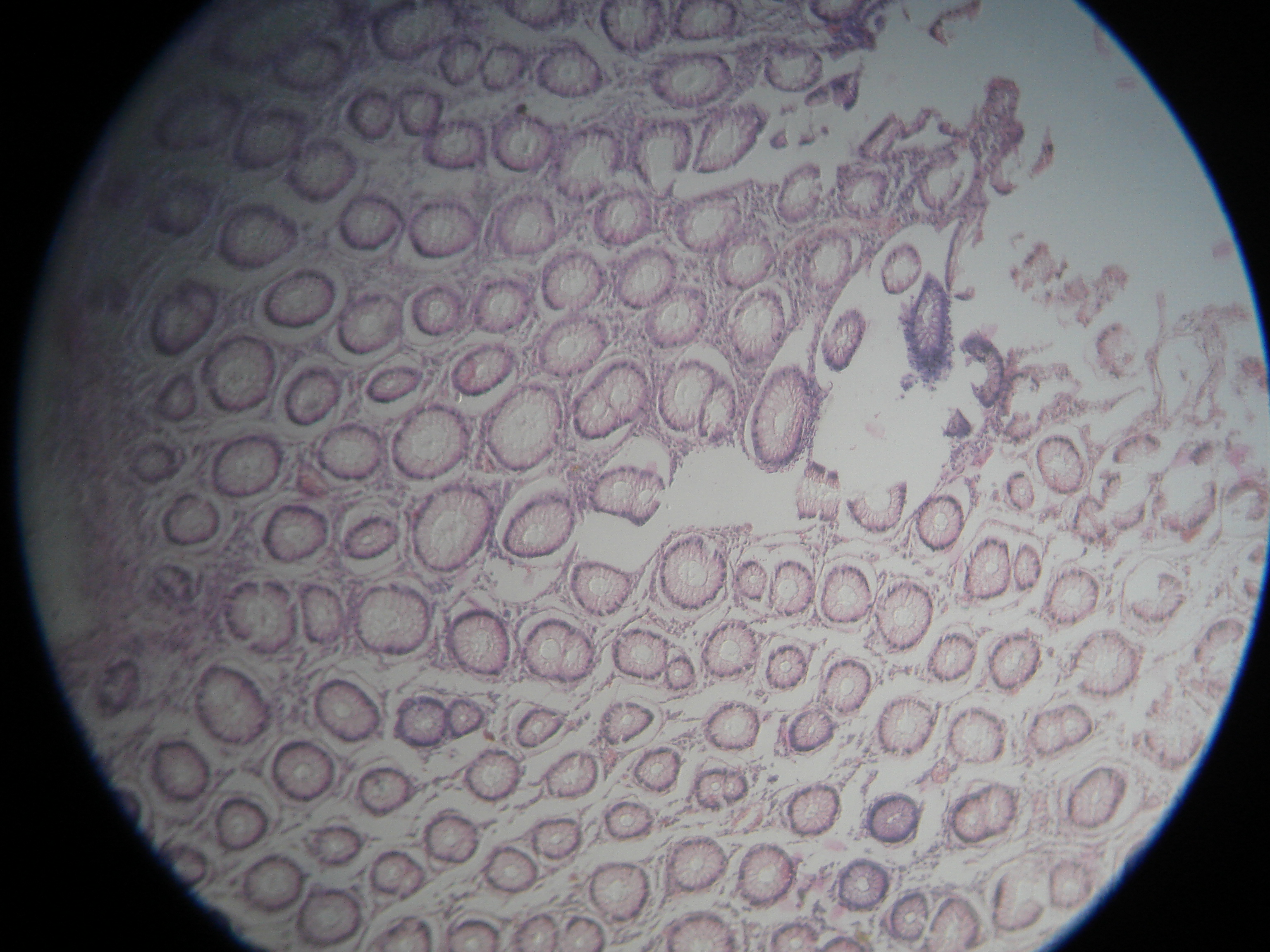
| Finding | Why it matters |
|---|---|
| Hyperplastic polyp | Often low risk when small and distal, but context and location matter. |
| Tubular adenoma | Precancerous; size, number and dysplasia determine surveillance. |
| Villous / tubulovillous adenoma | Higher-risk histology, especially when large or dysplastic. |
| Sessile serrated lesion | Important right-sided precursor pathway; complete removal and interval planning matter. |
| Invasive adenocarcinoma | Triggers staging, tumor markers and surgery/oncology planning. |
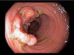
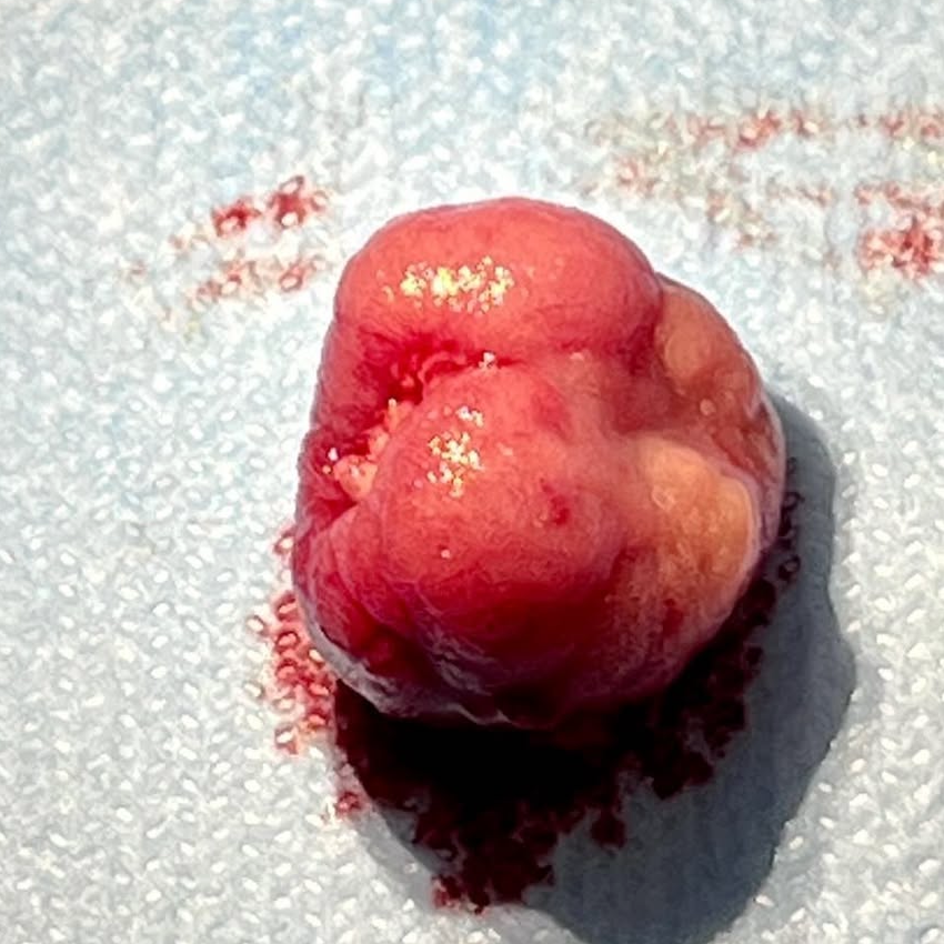
A removed polyp is prevention you can hold in a pot
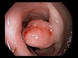
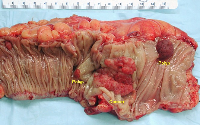
After colonoscopic removal, a polyp may look like a fleshy, bleeding lump rather than a neat textbook diagram. The important questions are whether it was removed completely, whether it was retrieved, whether removal was en bloc or piecemeal, its size and location, and what histology shows. Many colorectal cancers begin in adenomas or serrated lesions, so removing significant polyps early is the part of colonoscopy that can prevent cancer rather than merely detect it.
Follow-up depends on the worst polyp, the number of polyps, size and confidence of removal
Surveillance intervals below are broad, patient-facing examples from modern post-polypectomy guidance, especially US Multi-Society Task Force style intervals. Guidelines differ between countries and health systems, and this is not personal medical advice. Always ask your clinician what interval applies to your actual colonoscopy report, pathology report, bowel prep quality, family history and symptoms.
These examples assume a high-quality complete colonoscopy, adequate bowel prep, cecal intubation, complete removal and a patient without inflammatory bowel disease, hereditary syndrome or previous colorectal cancer. Poor prep, piecemeal removal, uncertainty about margins, strong family history or many polyps can shorten the interval.
| Pathology / finding | Usual meaning | Typical follow-up logic |
|---|---|---|
| Small distal hyperplastic polyps | Often not meaningfully precancerous when tiny and in the rectum/sigmoid. | Often treated like a normal colonoscopy, commonly 10 years in average-risk US-style guidance. |
| 1-2 small tubular adenomas under 10 mm | Low-risk precancerous adenomas if completely removed. | Often 7-10 years now, not automatically 3 years. |
| 3-4 small tubular adenomas under 10 mm | Higher polyp burden, but many patients no longer need an automatic 3-year interval. | Often 3-5 years; 5 years may be reasonable when prep, removal and pathology confidence are good. |
| 5-10 adenomas, any adenoma 10 mm or larger, villous/tubulovillous histology, or high-grade dysplasia | Higher-risk adenoma pattern or more advanced precancerous change. | Often 3 years if completely removed at a high-quality exam. |
| More than 10 adenomas | Raises concern for a polyposis tendency and missed synchronous lesions. | Often 1 year, plus consideration of genetic assessment. |
| 1-2 small sessile serrated lesions under 10 mm | Right-sided serrated pathway precursor; can be subtle and flat. | Often 5-10 years; shorter end if pathology distinction, prep or complete removal is uncertain. |
| 3-4 small sessile serrated lesions | Greater serrated burden. | Often 3-5 years. |
| Sessile serrated lesion 10 mm or larger, serrated lesion with dysplasia, or traditional serrated adenoma | Higher-risk serrated pathway lesion. | Often 3 years after confident complete removal. |
| Large sessile or laterally spreading lesion removed piecemeal, especially 20 mm or larger | Main worry is residual tissue at the scar rather than a new distant polyp. | Often an early site check around 6 months, then further surveillance such as 12 months after that depending on findings and local guidance. |
| Malignant polyp / invasive adenocarcinoma | Cancer has entered the polyp; depth, margins, lymphovascular invasion and grade decide next steps. | Needs specialist review. Some are cured endoscopically; others need colectomy, staging scans and oncology planning. |
The trend matters: for many low-risk findings the recommended interval has become longer, often 5, 7 or 10 years rather than reflexively 3. The exception is when the first exam was compromised, the lesion was large or piecemeal, dysplasia was advanced, there were many polyps, or the pathology suggests a higher-risk serrated or villous pathway.
FIT/FOBT sits beside this, not above it. If a person is returned to routine population screening after a normal or low-risk colonoscopy, they may resume annual FIT in some US-style pathways or biennial FIT in organized programs such as Australia or the NHS. But a negative FIT should not be used to postpone a surveillance colonoscopy that is due because of high-risk adenomas, advanced serrated lesions, piecemeal removal, poor prep or symptoms. A positive FIT/FOBT after any prior scope still needs proper clinical review and usually colonoscopy.
Inherited and tumor genetics
Inherited risk and tumor biomarkers answer different questions.
Inherited / germline genetics
Lynch syndrome genes, APC/FAP, MUTYH, POLE/POLD1, SMAD4, BMPR1A, STK11 and PTEN can change screening age, colonoscopy interval, surgical choices and family testing. Clues include young age, many polyps, multiple relatives, endometrial cancer patterns or rare hamartomatous polyps.
Tumor biomarkers
Mismatch repair/MSI testing is important for nearly all colorectal cancers. RAS, BRAF V600E, HER2 amplification, NTRK fusions and sometimes tumor mutation burden can guide metastatic treatment. CEA helps follow known cancer but is not a screening test.
If cancer is found
Detection shifts into staging and treatment planning.
Confirm tissue
Biopsy or resected polyp histology confirms cancer and depth of invasion.
Locate and mark
Colonoscopy report, distance from anal verge for rectal lesions and tattooing help surgical planning.
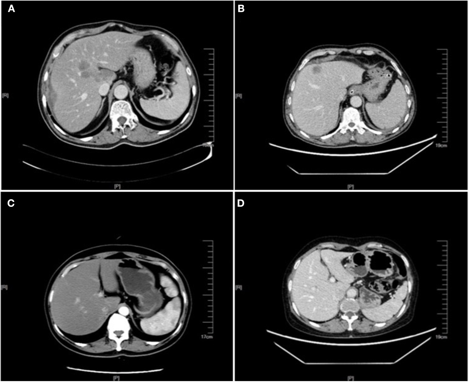
Stage
CT chest/abdomen/pelvis is common to look for spread, especially liver and lung metastases. Rectal cancer usually also needs pelvic MRI and sometimes endorectal ultrasound.
Baseline labs
CBC, iron studies, liver/kidney tests and CEA are used for safety, prognosis and future comparison.

Frailty changes the value of finding a lesion
For an older or frail person who cannot tolerate colonoscopy, CT colonography may answer an important question with less procedural burden. But detection is only useful if the result can lead to a benefit the person could actually live to enjoy: safer endoscopic treatment, elective surgery, stenting, radiotherapy, symptom relief, palliative planning or a decision not to intervene. Finding a cancer in someone unlikely to survive colectomy or chemotherapy may create anxiety and procedures without improving life.
False positives and overinvestigation
Colon cancer screening has strong logic, but the follow-up burden is real.
FOBT/FIT can be positive from benign bleeding, hemorrhoids, inflammation, ulcers or polyps. Colonoscopy can cause bleeding, perforation, sedation complications, cost, time off work and anxiety. The reason colorectal screening remains compelling is that it can prevent cancer by removing precursor lesions.
New blood cancer tests
Multi-cancer blood tests could help access, but they could also become a curse.
Liquid biopsy is useful once colorectal cancer is known, especially for tumor profiling, minimal residual disease research, recurrence monitoring and resistance testing. Multi-cancer early detection blood tests for asymptomatic people are different. PATHFINDER found more false-positive cancer signals than true cancers among positive tests. The NHS-Galleri randomized trial has now reported no lower incidence rate of stage III or IV cancers across 12 prespecified cancers after three screening rounds, compared with usual care alone.
For colon cancer specifically, proven pathways remain FOBT/FIT, stool DNA-FIT where appropriate, and colonoscopy follow-up. A blood test that creates an unclear cancer signal can lead to CT, PET/CT, endoscopy and anxiety without the preventive advantage of polyp removal.
The strongest argument for MCED is not colon cancer, breast cancer or cervical cancer, where established screening already exists. It is the hope of catching cancers that usually have no simple screening test and often present late: pancreatic cancer, ovarian cancer, liver cancer outside known cirrhosis surveillance, kidney cancer, sarcoma, some brain and CNS tumors, and other rare or anatomically hidden cancers. That hope is rational. The unresolved question is whether earlier signal detection in average-risk people leads to longer life or just more uncertain investigations.
The hard question is not whether a new test can find some cancers. It is whether using it at scale prevents deaths, finds treatable disease early enough, avoids too many false alarms, and does not overwhelm endoscopy, imaging and biopsy services. A convenient test with poor precursor detection may feel modern while quietly missing the main opportunity: preventing colon cancer before it exists.
A September 2026 Lancet commentary framed this as a "holy grail or cautionary tale" problem: hope and market pressure can move faster than mortality evidence, while false positives, false reassurance, overdiagnosis and downstream costs land on patients and health systems. That caution belongs on every new detection page, not just colon cancer.
Full-body MRI and direct-to-consumer screening
More imaging is not automatically more health.
Whole-body MRI has an understandable appeal: no ionizing radiation, one appointment, and the chance of finding a silent kidney cancer, brain tumor, sarcoma or aneurysm. For an individual paying privately, that may feel worth it, especially with strong anxiety, family history or a prior cancer experience. But it is different from a proven population screening program.
The appeal is especially strong for diseases people fear will otherwise be missed until too late: pancreatic cancer, brain tumors, ovarian cancer, kidney cancer, lymphoma, sarcoma, aneurysms or deep abdominal masses. A scan can sometimes find a real, important abnormality before symptoms. The problem is that the same scan can also find cysts, nodules, benign tumors, degenerative spine disease and borderline findings that look ominous but never would have harmed the person.
A celebrity example makes the dilemma easier to understand: Kim Kardashian publicly discussed a small brain aneurysm found on MRI. That kind of finding can be genuinely useful if it leads to expert review, blood-pressure control, smoking avoidance, stimulant/cocaine avoidance, family-history assessment and interval MRA/MRI follow-up when appropriate. It can also leave a person living with a frightening label for years when the aneurysm is too small or too low-risk to operate on safely. A broader detection guide belongs at DetectAneurysm.com, with deeper cerebral aneurysm detail at IntracranialAneurysm.com.
Aneurysms are not one disease. Cerebral aneurysms raise questions about MRA/CTA follow-up, rupture risk and sudden thunderclap headache. Aortic aneurysms divide into thoracic aortic aneurysm, abdominal aortic aneurysm and acute dissection or leaking/rupture pathways. AAA detection is more straightforward than many incidental MRI findings because abdominal ultrasound is quick, cheap, non-invasive and can identify a dangerous aorta before rupture, when elective repair is much safer than emergency surgery.
The American College of Radiology says there is not enough evidence to recommend total-body MRI screening for people with no symptoms, risk factors or family history suggesting disease. Recent reviews report that most asymptomatic people have at least one radiologic abnormality, many need extra tests, and only a small minority have a pathologically confirmed cancer. That means the lived result is often not reassurance but a queue of follow-up ultrasound, CT, targeted MRI, blood tests, biopsy or surveillance scans.
Insurance may cover targeted MRI for symptoms, known cancer staging, high-risk genetic syndromes such as Li-Fraumeni, or a defined clinical question. It often does not cover elective whole-body MRI for an average-risk, asymptomatic person. That does not forbid an informed private choice; it means the person should know that the scan may uncover uncertainty rather than health, and may not replace colonoscopy, FIT, mammography, cervical HPV testing, low-dose lung CT in eligible smokers or other proven site-specific screening.
Available now and in development
Specimen type matters: stool, blood, urine, sputum and saliva do different jobs.
For this colon page, stool and colonoscopy remain the main detection tools. Across the wider detection cluster, other specimens become relevant: urine for bladder and prostate triage, cervical/vaginal self-sampling for HPV, sputum or breath research for lung cancer, and saliva/oral rinse research for HPV-related throat and oral cancers.
Safe crosslinks
Related sites in the same medical cluster.
These links are included where they genuinely help the reader move from detection to treatment, age-based checks, specialist context or general cancer reference.
Evidence and reviews
Good starting points for screening and disease context.
CDC colorectal cancer screeningPractical screening options and follow-up after abnormal stool tests.
NCI colorectal cancerPatient and professional information on colon and rectal cancer.
ACS colorectal statistics 2026Current data on colorectal cancer incidence, mortality and rising younger-onset disease.
NCI younger-onset CRCDiet, microbiome, inflammation, obesity and lifestyle hypotheses for rising cancers under 50.
NCI warning signs in younger adultsRectal bleeding, anemia, diarrhea and abdominal pain should not be dismissed because of young age.
ACS colorectal risk factorsObesity, red and processed meat, smoking, alcohol, inactivity, family history and inherited syndromes.
NICE IBS guidanceIBS diagnosis should include assessment for cancer and inflammatory red flags.
NIDDK IBS symptomsPatient information on abdominal pain and bowel-habit change in IBS.
ACG ulcerative colitis guidelineColorectal cancer prevention and surveillance colonoscopy in UC beyond the rectum.
AGA IBD dysplasia surveillanceRisk-based colonoscopy surveillance for dysplasia in colonic inflammatory bowel disease.
BMJ clinical practice guidelineFIT, sigmoidoscopy and colonoscopy screening benefit/harm estimates.
NEJM IARC screening perspectiveReview of colorectal cancer screening evidence and program considerations.
Lancet colorectal cancer reviewRecent general review of colorectal cancer biology, diagnosis and treatment.
NEJM stool DNA-FIT trialNext-generation multitarget stool DNA screening performance.
Gross colectomy specimen imageWikimedia Commons image of opened colectomy specimen with invasive colorectal carcinoma and adenomatous polyps.
NEJM/IARC screening perspectiveMortality and incidence evidence across stool blood tests, FIT, sigmoidoscopy and colonoscopy.
JAMA USPSTF evidence reviewComparison of colorectal screening tests by mortality evidence, detection performance and harms.
USPSTF screening optionsUS recommendation listing FIT, stool DNA-FIT, CT colonography, flexible sigmoidoscopy and colonoscopy options.
Colonoscopy quality indicatorsCecal intubation, bowel prep quality, withdrawal time and adenoma detection as key quality measures.
Annals AI colonoscopy meta-analysisSystematic review finding higher ADR and adenomas per colonoscopy with computer-aided detection.
Lancet AI de-skilling studyObservational study of lower non-AI ADR after exposure to AI-assisted colonoscopy tools.
ACG summary: AI and de-skillingClinical commentary on the colonoscopy AI de-skilling signal and its limitations.
Cancer Council Australia colonoscopyAustralian patient information on full colonoscopy and bowel screening follow-up.
FDA Shield blood test approvalBlood cfDNA colorectal screening indication and performance summary.
NCI on Shield blood testPlain-English context for blood-based colorectal cancer screening.
NHS England bowel screeningFree FIT program age range and pathway.
Australia bowel screeningFree national program ages 45-74.
JAMA Network Open follow-up gapWhy timely colonoscopy after a positive stool test is a critical screening gap.
PATHFINDER MCED studyMulti-cancer blood-test positives included substantial false-positive follow-up.
NEJM NHS-Galleri trialFull randomized trial report on late-stage cancer diagnosis after three MCED screening rounds.
Lancet MCED cautionary taleWeinberg and Bretthauer's September 2026 critique of MCED evidence, harms and policy pressure.
ACR total-body MRI statementRadiology society statement on insufficient evidence for asymptomatic total-body MRI screening.
Whole-body MRI cancer screening reviewRecent review summarizing abnormal findings, follow-up investigations and confirmed cancer yield.
Kim Kardashian aneurysm reportPublic example of an MRI-detected small brain aneurysm and the anxiety/management questions it raises.
DetectAneurysm.comPlanned broader detection site for cerebral aneurysm, thoracic aortic aneurysm, abdominal aortic aneurysm, dissection, leak and rupture warning signs.
IntracranialAneurysm.comRelated patient-facing site for aneurysm risk, monitoring and treatment decisions.
NINDS cerebral aneurysm guideRisk factors, monitoring and treatment considerations for unruptured brain aneurysms.
ClinicalTrials.gov detection trialsCurrent colorectal screening and early-detection studies.