many skin cancers can be seen early.
Symptoms, risk, screening and diagnostic confirmation
Detect Skin Cancer is about choosing the right test for the right risk.
A skin cancer detection guide covering basal cell carcinoma, squamous cell carcinoma, melanoma, actinic keratoses, non-healing sores, dermoscopy and biopsy.
confirms subtype.
can spread, especially high-risk sites.
rarely spreads but can destroy local tissue.
Brief outline
What skin cancer means.
Skin cancer includes basal cell carcinoma, squamous cell carcinoma, melanoma and rarer tumors. Most are visible, which makes early detection possible.
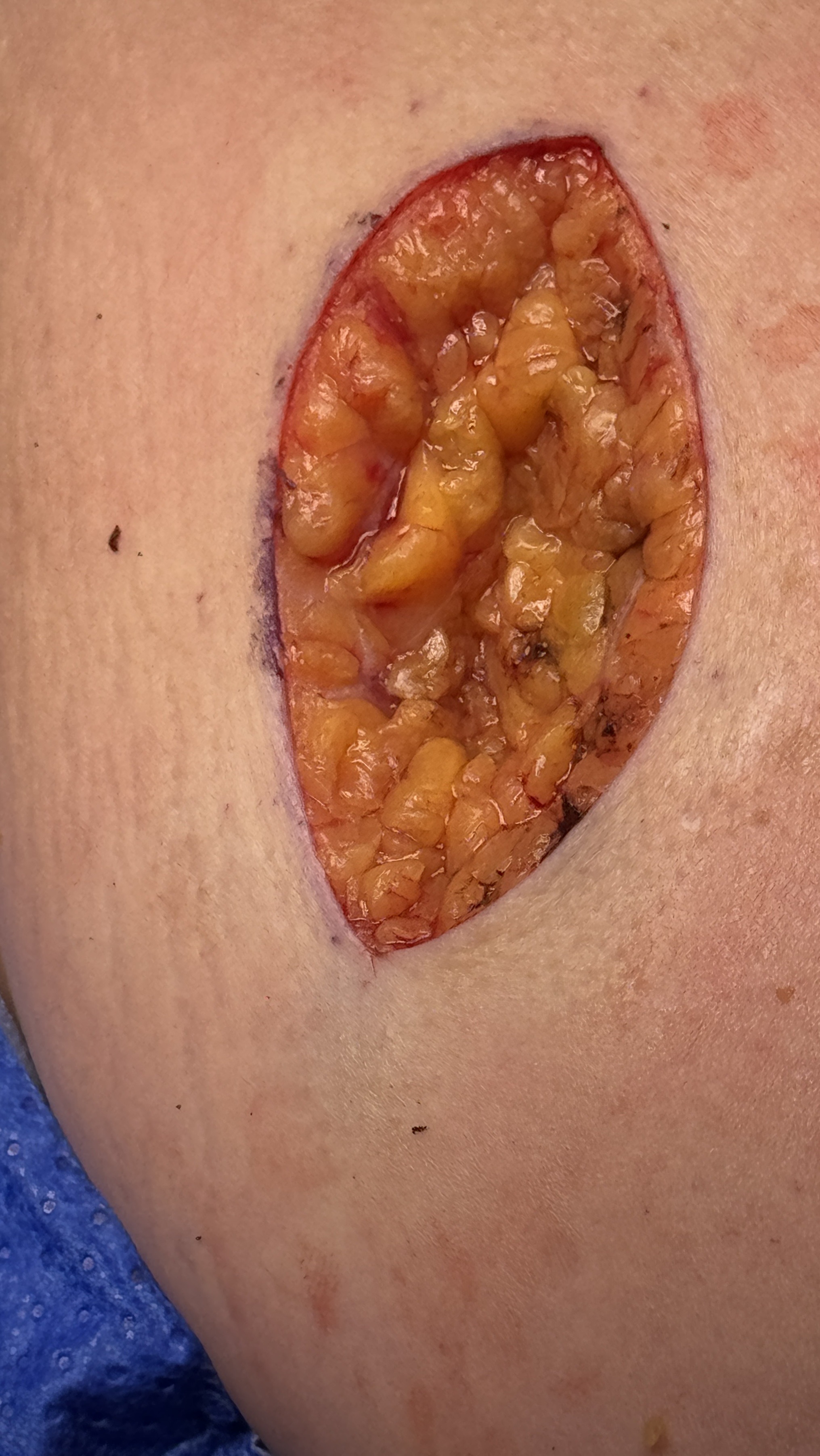
Detection frame
Screening is for people without symptoms. Diagnosis is for symptoms, abnormal examination, family history, imaging findings or abnormal labs. The same test can be sensible in one setting and misleading in another.
Do not ignore
Symptoms and signs not to ignore.
Risk and genetics
Demographics, risks and genetics.
Screening
Who should actually be screened?
Routine population screening varies by country. High-risk people often need regular skin surveillance.
Full skin examination
Checks visible and hard-to-see areas.
Dermoscopy
Improves accuracy for trained clinicians.
Shave, punch or excision biopsy
Choice depends on suspected cancer and site.
Margins and risk features
Depth, perineural invasion and differentiation guide care.
Nodal ultrasound, imaging or sentinel node biopsy
Reserved for selected high-risk SCC/Merkel/melanoma contexts, not routine BCC.
Photo-only reassurance
Images help monitoring but cannot replace biopsy when suspicion is high.
Images, labs and tissue
Useful tests and what they can miss.
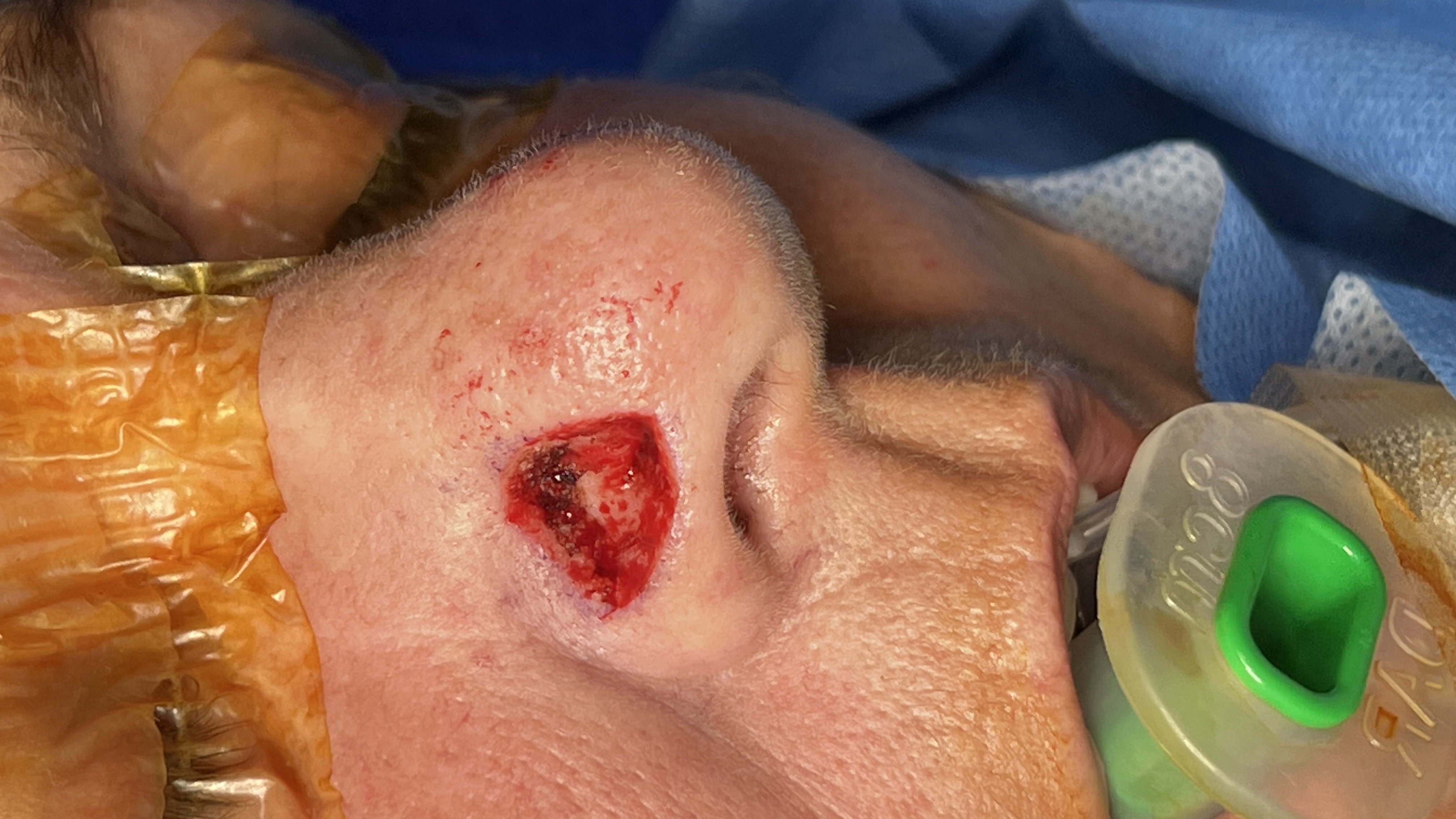
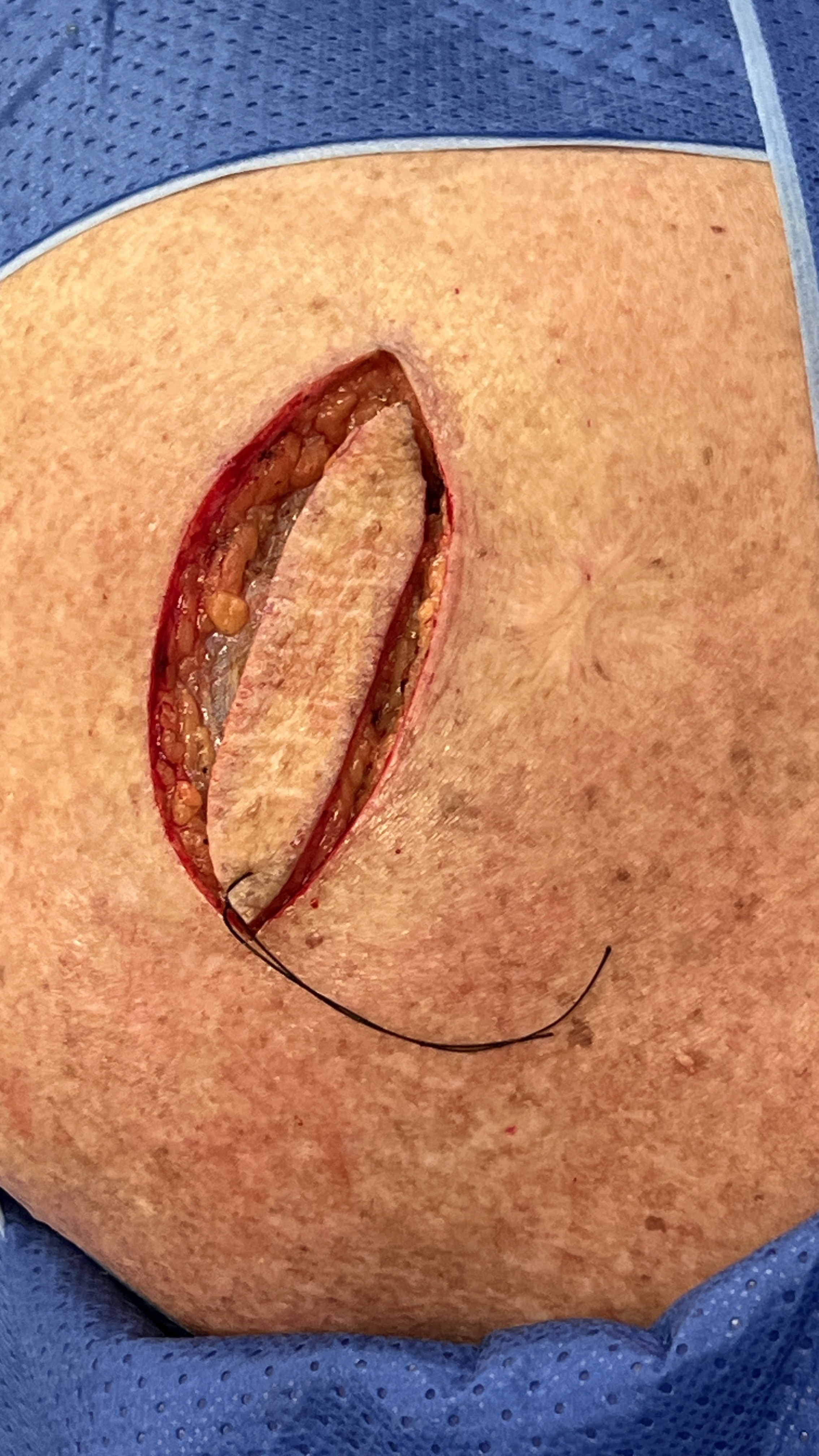
Detection usually needs a chain: suspicion, imaging or lab triage, tissue or cytology when needed, then pathology and molecular testing where it changes management.
Pros, cons and controversy
Benefits, harms and the false-positive problem.
The benefit is obvious because lesions are accessible. The harm is excessive biopsies of harmless spots, but the bigger danger is ignoring a persistent non-healing lesion.
Sources and trials