large soft-tissue lumps deserve imaging.
Symptoms, risk, screening and diagnostic confirmation
Detect Sarcoma is about choosing the right test for the right risk.
A sarcoma detection guide covering soft-tissue lumps, retroperitoneal tumors, MRI, CT, core biopsy and referral before surgery.
deep or fixed masses are concerning.
best local soft-tissue map.
should be planned before surgery.
Brief outline
What sarcoma means.
Sarcomas are rare cancers of connective tissue, bone, muscle, fat, vessels or other mesenchymal tissues. The main detection rule is not to remove a suspicious lump casually before imaging and planned biopsy.
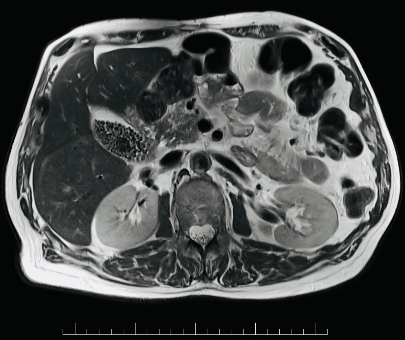
Detection frame
Screening is for people without symptoms. Diagnosis is for symptoms, abnormal examination, family history, imaging findings or abnormal labs. The same test can be sensible in one setting and misleading in another.
Do not ignore
Symptoms and signs not to ignore.
Risk and genetics
Demographics, risks and genetics.
Screening
Who should actually be screened?
There is no general sarcoma screening. In genetic syndromes such as Li-Fraumeni, whole-body MRI surveillance may be used.
MRI with contrast
Preferred for limb/trunk soft-tissue masses.
CT abdomen/chest
Useful for retroperitoneal disease and lung staging.
Core needle biopsy
Planned through a route that will not compromise surgery.
IHC and molecular tests
Sarcoma subtype often needs specialist pathology.
Chest CT/PET selected
Lung metastases are important in many sarcomas.
Unplanned excision
Removing a suspicious lump before imaging can make definitive surgery harder.
Images, labs and tissue
Useful tests and what they can miss.
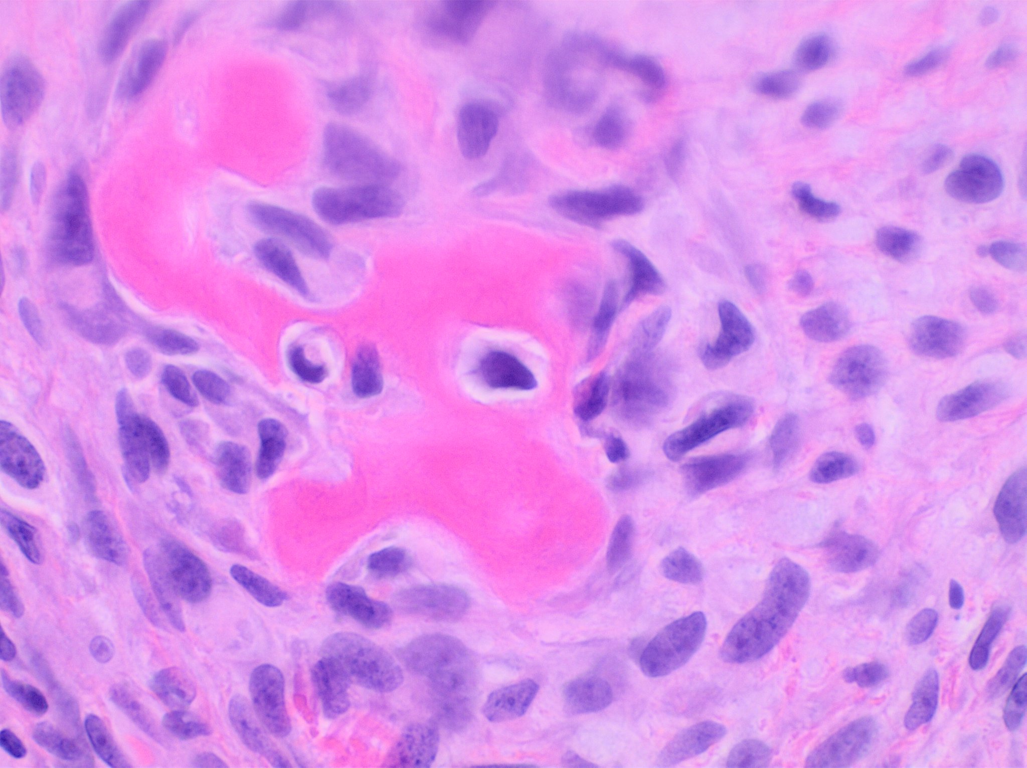
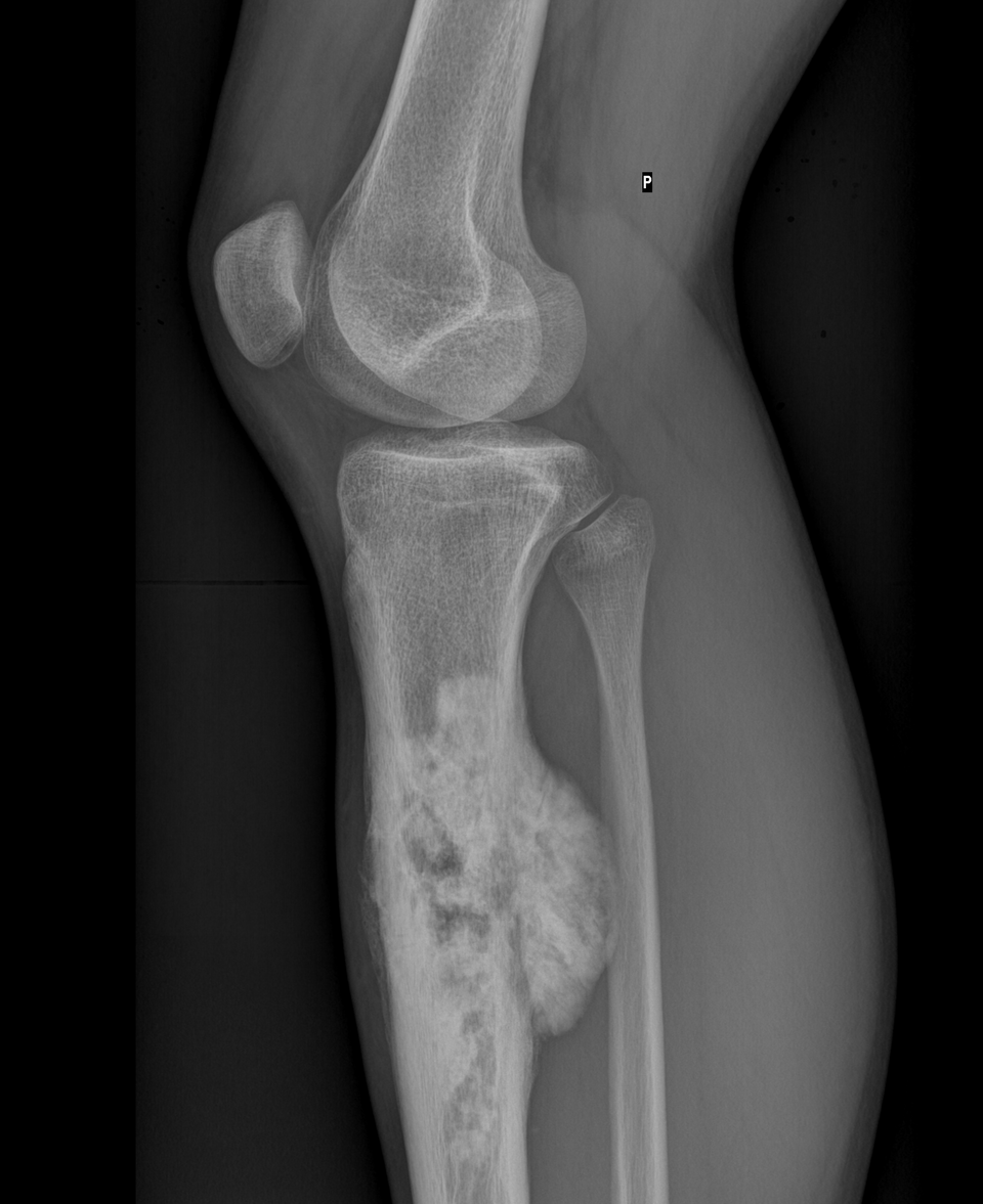
Detection usually needs a chain: suspicion, imaging or lab triage, tissue or cytology when needed, then pathology and molecular testing where it changes management.
Pros, cons and controversy
Benefits, harms and the false-positive problem.
The avoidable harm is a 'whoops' operation: a sarcoma shelled out as a presumed lipoma, contaminating tissue planes and complicating cure.
Sources and trials