persistent ulcers need review.
Symptoms, risk, screening and diagnostic confirmation
Detect Oral Cancer is about choosing the right test for the right risk.
An oral cancer detection guide covering tongue lesions, mouth ulcers, red/white patches, HPV-related disease, examination, imaging and biopsy.
confirms dysplasia or cancer.
more relevant to oropharynx than most oral cavity cancers.
can be first sign of spread.
Brief outline
What oral cancer means.
Oral cancer is often squamous cell carcinoma of the tongue, floor of mouth, gums or other mucosa. Persistent lesions need direct examination and biopsy.

Detection frame
Screening is for people without symptoms. Diagnosis is for symptoms, abnormal examination, family history, imaging findings or abnormal labs. The same test can be sensible in one setting and misleading in another.
Do not ignore
Symptoms and signs not to ignore.
Risk and genetics
Demographics, risks and genetics.
Screening
Who should actually be screened?
No broad oral cancer screening program exists for average-risk adults, but dental and medical examinations can catch visible lesions.
Oral inspection and palpation
Good lighting, tongue movement and neck palpation matter.
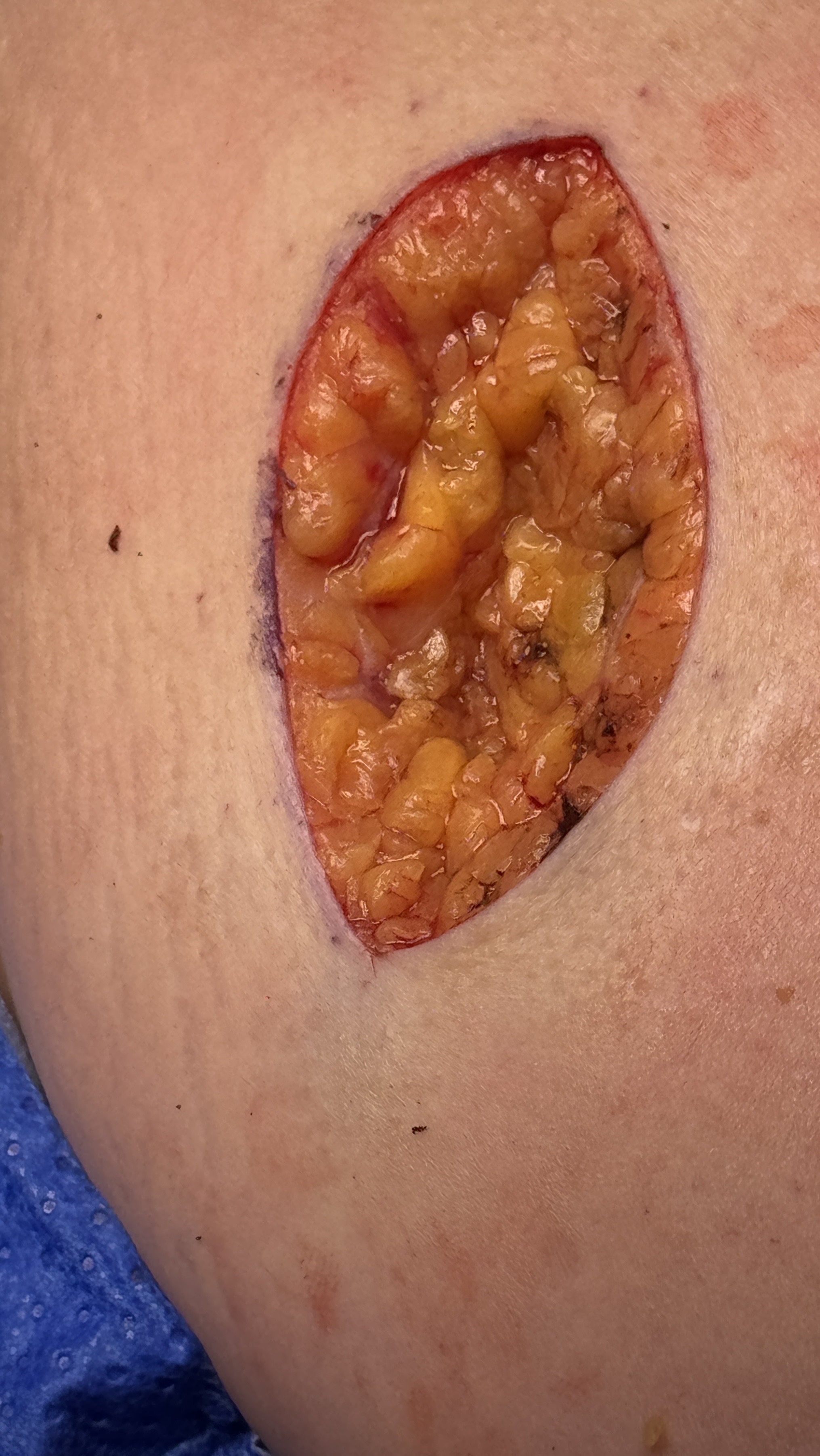
Biopsy
Confirms dysplasia, SCC or benign disease.
CT/MRI
Maps deeper invasion.
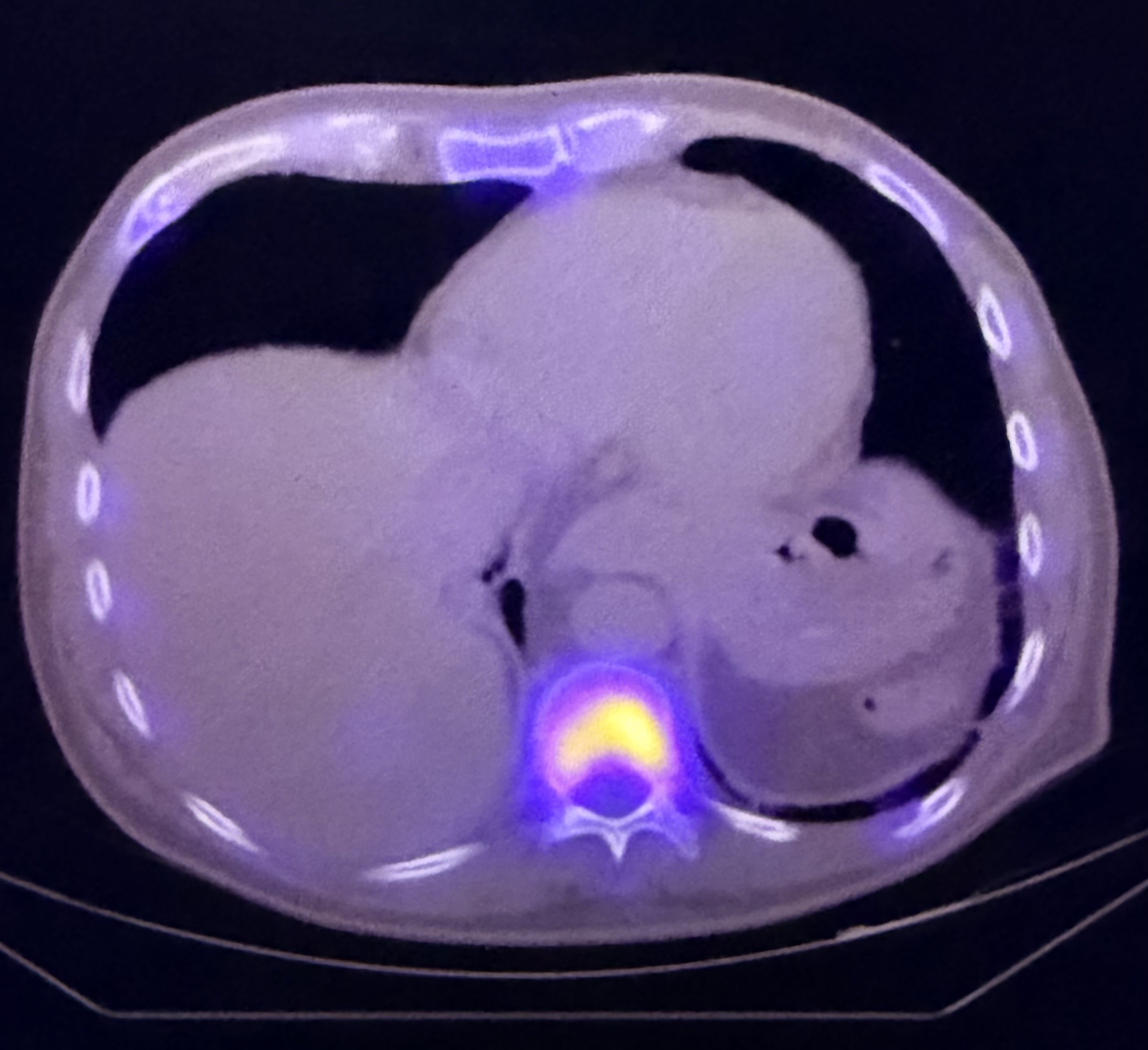
PET-CT
Selected staging for advanced disease.
p16/HPV context
More central in oropharyngeal cancer.
Mouthwash or antibiotics
Should not delay biopsy of persistent lesions.
Images, labs and tissue
Useful tests and what they can miss.
Detection usually needs a chain: suspicion, imaging or lab triage, tissue or cytology when needed, then pathology and molecular testing where it changes management.
Pros, cons and controversy
Benefits, harms and the false-positive problem.
The risk is under-investigation: ulcers are common, but persistence is the line. Adjunctive dyes/lights cannot replace biopsy.
Sources and trials