calcium, renal, anemia and bone lesions.
Symptoms, risk, screening and diagnostic confirmation
Detect Myeloma is about choosing the right test for the right risk.
A myeloma detection guide covering CRAB features, SPEP, immunofixation, serum free light chains, urine tests, bone marrow biopsy and whole-body imaging.
detects many monoclonal proteins.
catch light-chain disease.
low-dose whole-body CT/PET/MRI can reveal lytic lesions.
Brief outline
What myeloma means.
Multiple myeloma is a plasma-cell cancer. It can cause anemia, kidney injury, high calcium, bone pain, fractures and infections.
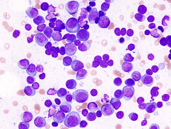
Detection frame
Screening is for people without symptoms. Diagnosis is for symptoms, abnormal examination, family history, imaging findings or abnormal labs. The same test can be sensible in one setting and misleading in another.
Do not ignore
Symptoms and signs not to ignore.
Risk and genetics
Demographics, risks and genetics.
Screening
Who should actually be screened?
There is no routine population screening program. MGUS or smoldering myeloma surveillance is different from screening the general public.
SPEP and immunofixation
Finds and types monoclonal protein.
Serum free light chains
Important for light-chain and non-secretory patterns.
UPEP/urine immunofixation
Can detect urinary light chains.
Bone marrow biopsy
Measures plasma-cell burden and genetics.
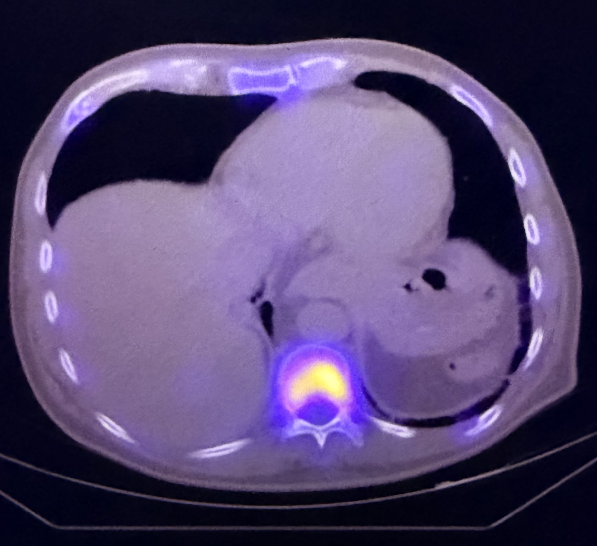
Whole-body low-dose CT/PET-CT/MRI
Assesses lytic lesions and focal marrow disease.
Borderline proteins
MGUS is common with age and not the same as myeloma.
Images, labs and tissue
Useful tests and what they can miss.
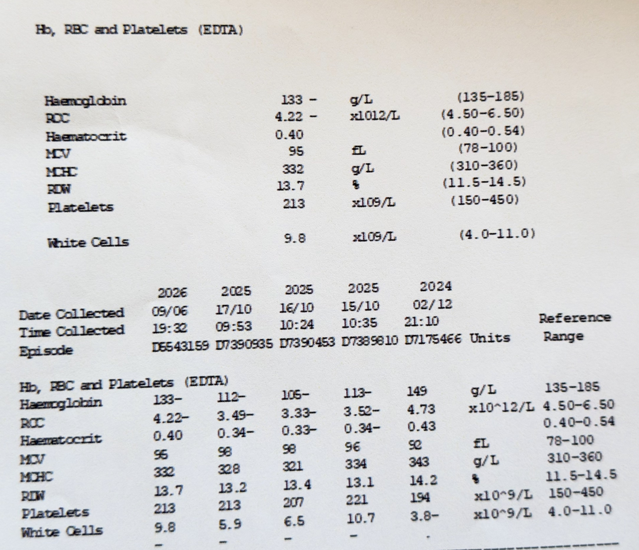
Detection usually needs a chain: suspicion, imaging or lab triage, tissue or cytology when needed, then pathology and molecular testing where it changes management.
Pros, cons and controversy
Benefits, harms and the false-positive problem.
Earlier recognition prevents renal failure and fractures, but finding MGUS creates lifelong surveillance for many people who never develop myeloma.
Sources and trials