many are found on scans for other reasons.
Symptoms, risk, screening and diagnostic confirmation
Detect Meningioma is about choosing the right test for the right risk.
A meningioma detection guide covering incidental MRI findings, seizures, vision change, focal deficits, CT/MRI, surveillance and why benign tumors can still kill.
defines size, edema and location.
some small tumors are monitored.
benign location can still be dangerous.
Brief outline
What meningioma means.
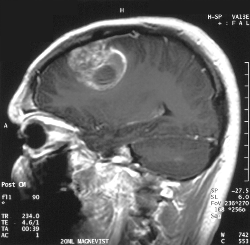
Meningiomas arise from meninges and are often benign, but location, size, edema, venous sinus invasion or higher grade can make them dangerous.
Detection frame
Screening is for people without symptoms. Diagnosis is for symptoms, abnormal examination, family history, imaging findings or abnormal labs. The same test can be sensible in one setting and misleading in another.
Do not ignore
Symptoms and signs not to ignore.
Risk and genetics
Demographics, risks and genetics.
Screening
Who should actually be screened?
No population screening exists. Detection is symptom-led or incidental, with MRI surveillance based on size, growth and risk.
MRI brain with contrast
Best standard test for suspected meningioma.
CT brain
Often first in acute symptoms.
Serial MRI
Tracks growth in observed tumors.
Formal visual fields
Important for optic pathway lesions.
Surgery/biopsy pathology
Defines WHO grade when removed.
Incidentaloma anxiety
Many small meningiomas never need treatment.
Images, labs and tissue
Useful tests and what they can miss.
Detection usually needs a chain: suspicion, imaging or lab triage, tissue or cytology when needed, then pathology and molecular testing where it changes management.
Pros, cons and controversy
Benefits, harms and the false-positive problem.
The phrase benign can be misleading. Small stable tumors may be watched safely; tumors compressing brain, optic nerves or venous sinuses can threaten life or function.
Sources and trials