broad population screening is recommended for average-risk adults.
Hematuria, renal masses and incidental CT findings
Kidney cancer is often found by chance, but blood in urine must not be ignored.
A practical detection guide to hematuria, flank pain, ultrasound, CT renal mass protocols, MRI, biopsy, Bosniak cysts, hereditary syndromes and why average-risk population screening is not routine.
visible or persistent microscopic blood in urine needs evaluation.
renal mass protocols define enhancement, stage and surgical planning.
VHL, Birt-Hogg-Dube, HLRCC and other syndromes change surveillance.
Brief outline
Most kidney cancers are renal cell carcinomas.
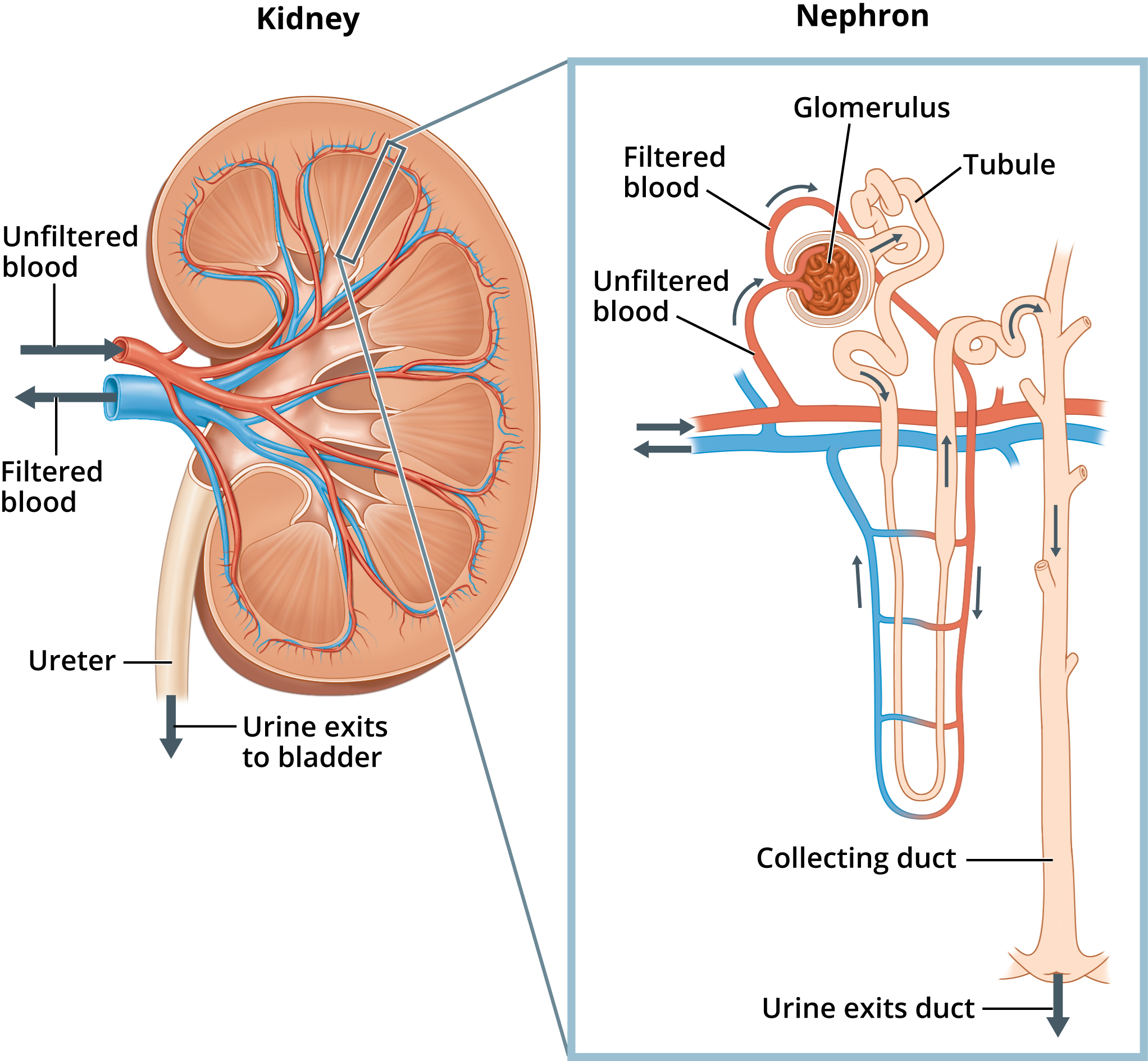
Renal cell carcinoma can be clear cell, papillary, chromophobe or rarer subtypes. Some cancers are small incidental renal masses; others present with hematuria, anemia, weight loss or metastatic symptoms.
Detection frame
Detection splits into two pathways: investigating hematuria or symptoms, and managing an incidental renal mass found on ultrasound, CT or MRI done for another reason.
Do not ignore
Hematuria is the headline warning sign.
Risk and genetics
Smoking, obesity, hypertension and inherited syndromes matter.
Screening
Average-risk screening is not routine.
Urine dipsticks and ultrasound are not proven population screening tools for average-risk adults. High-risk genetic syndromes are different: they may need scheduled MRI/CT/ultrasound surveillance in specialist programs.
Urinalysis and urine culture
Useful for hematuria workup but cannot rule out kidney cancer.
Ultrasound
Good for cysts and hydronephrosis, less definitive for complex or small solid masses.
CT renal mass protocol
Assesses enhancement, renal vein/IVC invasion, nodes and metastases.
MRI
Helpful for contrast allergy, equivocal CT, venous invasion and hereditary surveillance.
Renal mass biopsy
Can avoid unnecessary surgery in benign or metastatic/inflammatory mimics.
Urine cytology
More relevant to urothelial cancer than typical renal cell carcinoma.
Images, labs and tissue
Renal masses are characterized by enhancement and anatomy.
Ultrasound can find cystic versus solid masses. CT or MRI with renal mass protocol defines enhancement, fat, vessels, nodes and metastases. Biopsy is selective: useful when it changes management, but not required for every surgically treated mass.
Pros, cons and controversy
Finding small renal masses can help, but can also overtreat.
Many small renal masses grow slowly, especially in older or frail patients. Active surveillance, ablation, partial nephrectomy and radical nephrectomy all have roles. Whole-body MRI may find kidney masses but also benign cysts that trigger follow-up.
Sources and trials